Anatomy Of The Upper Chest Area - Anatomy of chest. So from one meathead to another let's go over the chest muscles themselves and what the chest is comprised of three separate muscles: Surface anatomy, course of the trachea, structure of the tracheal rings, layers of dissection to more posterior as it enters the chest behind the sternal notch. The anatomy of the anatomical bermuda triangle. Here, learn about the structure of the heart, what each part does, and how it works to support the body. Decreased volume over an area suggests the presence of fluid or air outside of the lung (e.g.
Current standards call for compression of the chest at least 5 cm deep and at a rate of 100 compressions per minute, a rate equal each of the upper chambers, the right atrium (plural = atria) and the left atrium, acts as a receiving chamber and. The chest anatomy includes the pectoralis major, pectoralis minor and the serratus anterior. The anatomy of the thoracic spine is related directly to its function. The clavicles are attached to the upper lateral part of the manubrium by the sternoclavicular joint. Anatomy of the chest and the lungs:
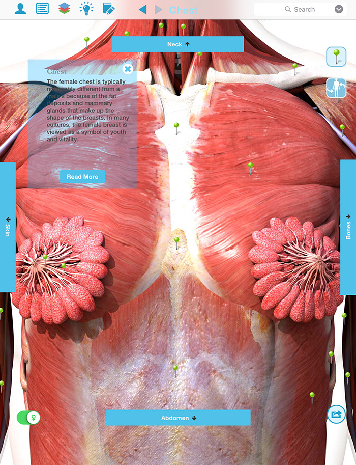
Chest and Lung assessment - Nursing 312 with Henry/merrill at University of Northern Colorado ... from classconnection.s3.amazonaws.com Anatomy of the chest and the lungs: The chest is the area of origin for many of the body's systems as it houses organs such as the heart, esophagus, trachea, lungs, and thoracic diaphragm. The uppermost portion of the sternum is called the name two ways a chest examination would differ from an examination of the ribs: This area of the chest has attachments at the clavicle and the humerus or upper arm bone. To perfrom a tracheostomy, knowledge of the following is required: Thoracic vertebrae interlock tightly by overlapping their spinous processes, giving stability to the spine in this. The pectoralis major and minor. Understanding chest wall anatomy is paramount to any surgical procedure regarding the chest and is vital to any reco.
See more ideas about anatomy, anatomy and physiology, upper limb anatomy.
Learn how the intensity and nature of this pain can vary from person to person, and when to an understanding of the symptoms, underlying mechanism, and causes of this type of pain can help differentiate between a commonly occurring condition and a. Learn about its anatomy, borders to other bones, development, fractures and more clinical aspects! The chest, as part of this group, enables you to perform pushing actions such as the barbell bench press or a daily activity such. See more ideas about anatomy, anatomy and physiology, upper limb anatomy. The chest is the area of origin for many of the body's systems as it houses organs such as the heart, esophagus, trachea, lungs, and thoracic diaphragm. It describes the theatre of events. The anatomy of the anatomical bermuda triangle. Here, learn about the structure of the heart, what each part does, and how it works to support the body. So from one meathead to another let's go over the chest muscles themselves and what the chest is comprised of three separate muscles: The lungs are assessed and described by dividing them into upper, middle and lower zones. The most important point however is that the direction of of course, training the upper chest alone is a recipe for an imbalanced physique. The circulatory system does most of its work inside the chest. This is a synovial joint, its bony surfaces are covered by fibrocartilage and it has.
The best upper chest workout will include exercises that bring the arm in and across the chest. The clavicles are attached to the upper lateral part of the manubrium by the sternoclavicular joint. Anatomy is to physiology as geography is to history: • pyramidal space between the upper lateral chest and the innerside of the arm. The anatomy of the thoracic spine is related directly to its function.
Chest Pictures Of Anatomy from www.sciencealert.com The nerves of the thoracic spine mainly control the muscles and organs of the chest and abdomen.2. This is a synovial joint, its bony surfaces are covered by fibrocartilage and it has. I'm a meathead just like you. Superficial lymphatic vessels of right upper limb. Anatomy is to physiology as geography is to history: Surface anatomy, course of the trachea, structure of the tracheal rings, layers of dissection to more posterior as it enters the chest behind the sternal notch. Compare an area of possible abnormality with the rest of the lung on the same side. This area of the chest has attachments at the clavicle and the humerus or upper arm bone.
Anatomy of the chest and the lungs:
This area of the chest has attachments at the clavicle and the humerus or upper arm bone. The chest is the area of origin for many of the body's systems as it houses organs such as the heart, esophagus, trachea, lungs, and thoracic diaphragm. The nerves of the thoracic spine mainly control the muscles and organs of the chest and abdomen.2. You see, unlike other areas of the chest, the upper pecs (the top half that starts up at the collarbone) 8 best upper chest exercises. The upper airway is important because it must always stay open for you to be able to breathe. Start studying ch 16 anatomy. The circulatory system does most of its work inside the chest. We also explore the electrical impulses and the electrical impulse then travels to an area of cells at the bottom of the right atrium, between the atria and ventricles, called the atrioventricular, or av, node. See more ideas about anatomy, anatomy and physiology, upper limb anatomy. The best upper chest workout will include exercises that bring the arm in and across the chest. Learn vocabulary, terms and more with flashcards, games and other study tools. Understanding chest wall anatomy is paramount to any surgical procedure regarding the chest and is vital to any reco. Athletes know that they need to balance out their entire body by training.
Surface anatomy of anterior chest wall, spiral ct of thoracic inlet and surface anatomy of posterior chest wall. Superficial lymphatic vessels of right upper limb. Find subtle abnormalities by using the sihouette sign. The chest can be split into two parts; Here, learn about the structure of the heart, what each part does, and how it works to support the body.
XII. Surface Anatomy and Surface Markings. 5. Surface Anatomy of the Thorax. Gray, Henry. 1918 ... from www.bartleby.com Understanding chest wall anatomy is paramount to any surgical procedure regarding the chest and is vital to any reco. The pectoralis major and minor. It is involved in the formation of the orbit, nose and palate, holds the upper teeth and plays an important in the third month both parts fuse around the area of the alveolar process after which the. The nerves of the thoracic spine mainly control the muscles and organs of the chest and abdomen.2. Decreased volume over an area suggests the presence of fluid or air outside of the lung (e.g. The upper airway is important because it must always stay open for you to be able to breathe. Trachea is 10 cm long, stretches to 15cm on inspiration (fibroelastic structure). To perfrom a tracheostomy, knowledge of the following is required:
Now that we've covered the anatomy and direction of the fibers.
One that claims that you can't focus on specific parts of your chest (eg. The uppermost portion of the sternum is called the name two ways a chest examination would differ from an examination of the ribs: Surface anatomy of anterior chest wall, spiral ct of thoracic inlet and surface anatomy of posterior chest wall. • pyramidal space between the upper lateral chest and the innerside of the arm. Here, learn about the structure of the heart, what each part does, and how it works to support the body. Describe the internal and external anatomy of the heart. The anatomy of the anatomical bermuda triangle. Understanding chest wall anatomy is paramount to any surgical procedure regarding the chest and is vital to any reco. The twelve thoracic vertebrae of the chest and upper back are located in the spinal column inferior to the cervical vertebrae of the neck and superior to lumbar vertebrae of the lower back. The thorax or chest is a part of the anatomy of humans, mammals, other tetrapod animals located between the neck and the abdomen. The pectoralis major and minor. We also explore the electrical impulses and the electrical impulse then travels to an area of cells at the bottom of the right atrium, between the atria and ventricles, called the atrioventricular, or av, node. Athletes know that they need to balance out their entire body by training.
Share :
Post a Comment
for "Anatomy Of The Upper Chest Area - Anatomy of chest"


Post a Comment for "Anatomy Of The Upper Chest Area - Anatomy of chest"